I'm fortunate enough to work with X-ray sources, so I wanted to see for myself which everyday materials react visibly. My goal was to find something common in a household that could potentially be paired with a photodiode to act as a DIY X-ray detector.
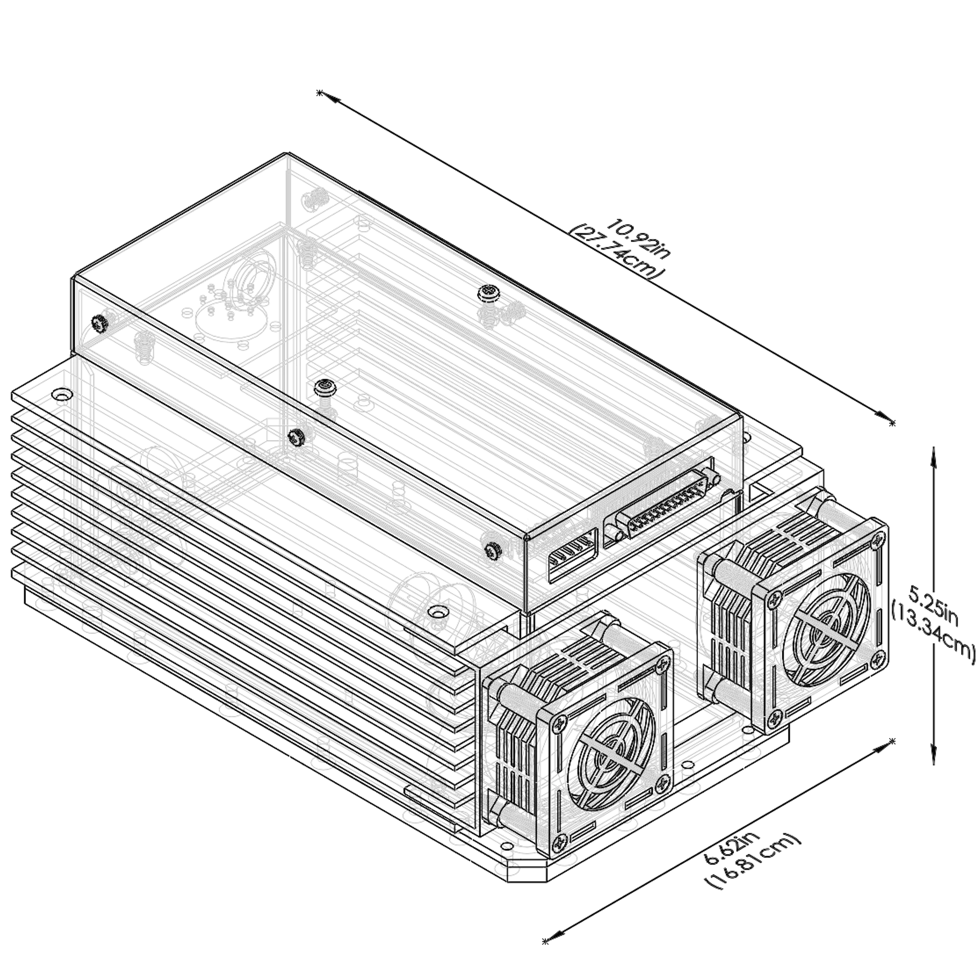
To illuminate the samples, I used an SB-80-1k source at full power: 80kV and 1mA anode current. This is roughly the same power level used in mammography. Both the source and the test materials were kept inside a shielded enclosure, safely away from me. You can find more details about the tube on the manufacturer's website:
sourceray.com — SB-80 series
sourceray.com — SB-80 series
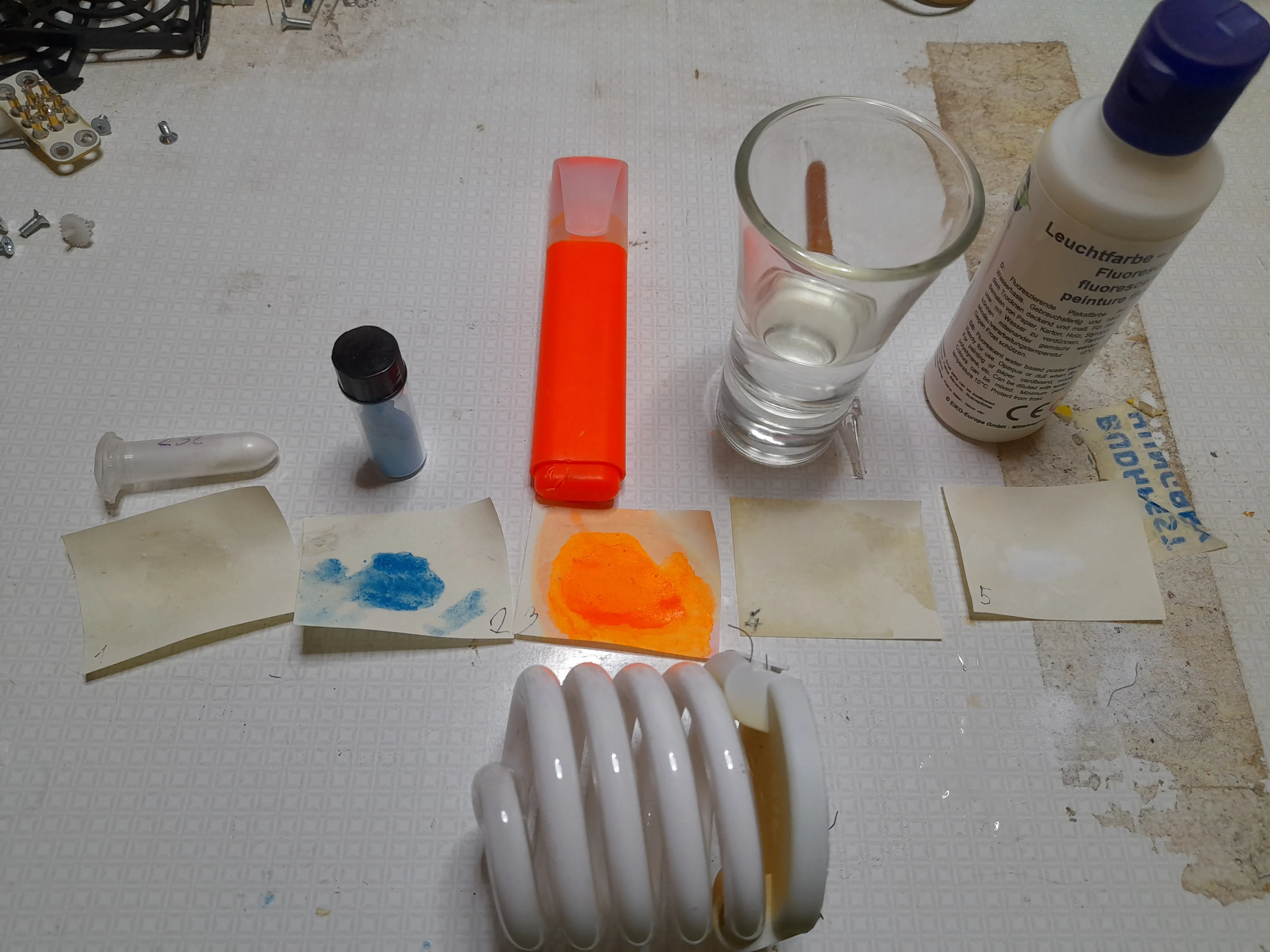
I started my testing with the following materials (from left to right):
- Strontium aluminate — glow-in-the-dark pigment (dopant unknown), glows green.
- Strontium aluminate — another glow pigment, this one glowing blue.
- Fluorescein — extracted from a highlighter pen.
- Quinine — from tonic water; dripped onto paper multiple times and dried to concentrate it.
- UV fluorescent paint.
- Bottom: A mercury-based compact fluorescent lamp (CFL) with its phosphor coating.
Let's start with the failures. The fluorescein, the quinine, and the UV fluorescent paint showed no reaction at all. I even tried placing banknotes in the beam path, but the red and blue fibers that normally glow under UV light didn't respond to X-rays. As a general takeaway, I found that materials designed for UV fluorescence usually don't react here.
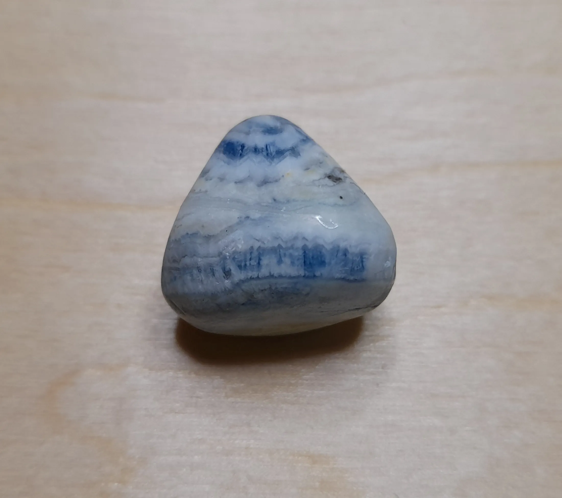
I also examined various minerals, but unfortunately, none of them were successful — not even Scheelite ($CaWO_4$), which is definitely a material used for X-ray detection. I'm certain the mineral I have is Scheelite and that I irradiated it properly; the lack of a visible glow might be due to impurities in the natural stone. Theoretically, 80kV should be enough to trigger a reaction, and I believe the 1mA intensity was sufficient as well, since I placed the samples directly at the X-ray exit window where the radiation is concentrated into a roughly 1cm² area. This made me wonder about the massive voltages and anode currents Röntgen must have used back in the day.
I tested the following minerals, ordered by their expected reaction intensity and typical colors. As I mentioned, I couldn't observe any light from them, likely because a higher tube voltage was required.
I tested the following minerals, ordered by their expected reaction intensity and typical colors. As I mentioned, I couldn't observe any light from them, likely because a higher tube voltage was required.
| Mineral | Expected Intensity | Typical Color |
|---|---|---|
| Scheelite | Excellent (very strong) | Light Blue / White |
| Fluorite | Strong | Violet / Blue / Green |
| Calcite | Medium / Strong | Orange / Red |
| Sodalite | Medium | Orange |
| Selenite | Weak | Pale Blue |
| Chalcedony | Very Weak | Pale Green (rare) |
Now for the things that actually worked. Generally, I noticed that materials referred to as "phosphors" (luminescent powders) tend to react well.
Strontium aluminate $SrAl_2O_4$: Both the blue and green versions reacted well. The green one seemed to have a longer afterglow — when I turn off the source, you can see the intensity drop, but the green luminescence persists even after I remove the shielding from the deactivated tube. (In the video, I turn the source on at 0:20 and off at 2:26.) The usual dopants of strontium aluminate are Europium (Eu), Dysprosium (Dy), or Boron (B).
A compact fluorescent lamp (CFL) contains mercury, which is excited by an electric field to create UV light inside the tube. This UV is then converted to white light by phosphors. The color temperature depends on the specific phosphor mix. I once had a very cheap lamp that glowed purple until it warmed up — it felt like it was making the room darker! In quality lamps, white light is produced by a "tri-phosphor" blend:
- Blue phosphor: Barium magnesium aluminate (BAM) — $BaMgAl_{10}O_{17}:Eu^{2+}$
- Green phosphor: Lanthanum phosphate or Terbium-magnesium aluminate — $LaPO_4:Ce^{3+}, Tb^{3+}$ or $CeMgAl_{11}O_{19}:Tb^{3+}$
- Red phosphor: Yttrium oxide — $Y_2O_3:Eu^{3+}$
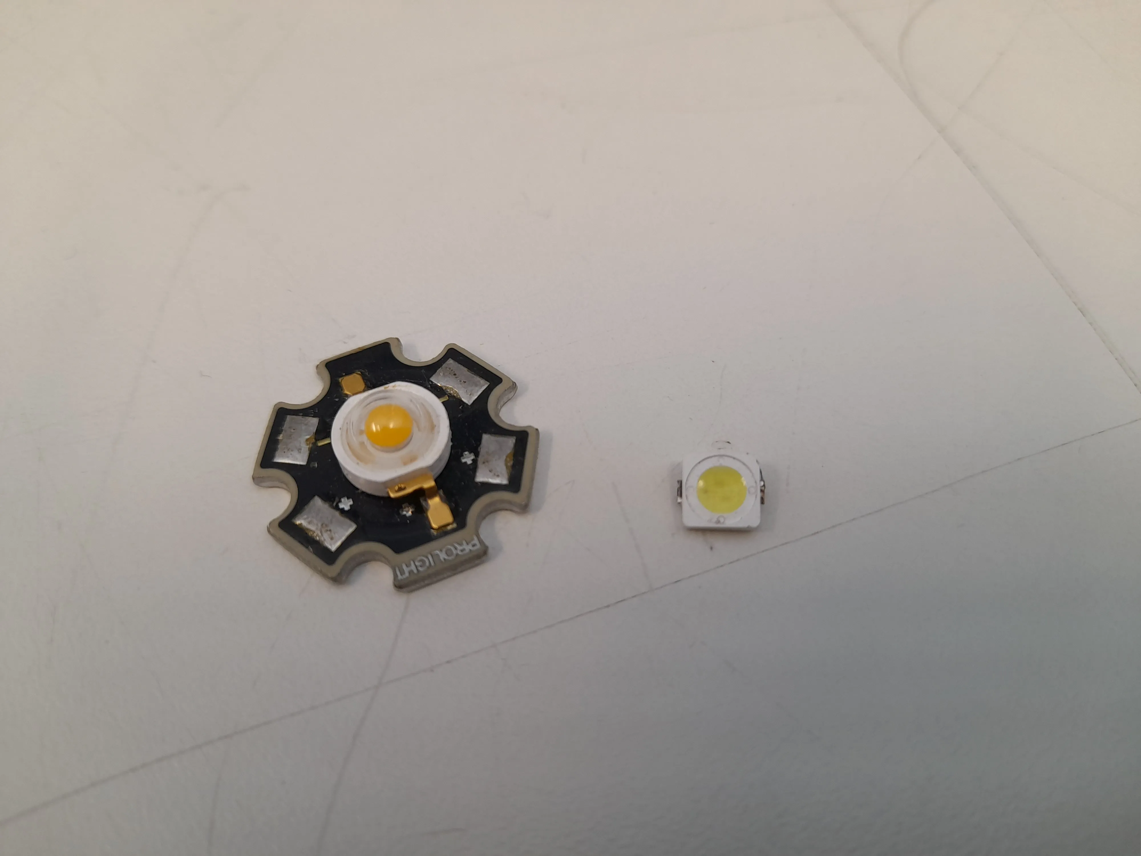
After the success with the CFL, I tried two types of white LEDs. These also use various phosphor mixtures to create white light.
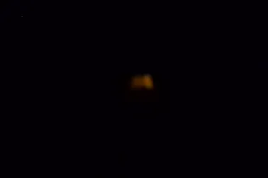
The larger, higher-power LED on the left glowed weakly with a yellowish-orange tint.
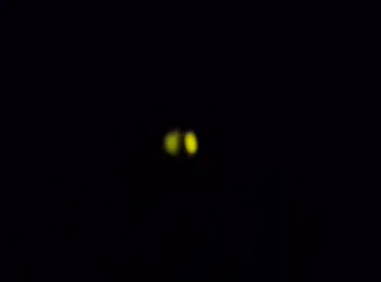
The smaller power LED was much brighter, emitting a greenish-yellow light. It seems that most white LEDs containing yellow phosphor will react to X-ray radiation.
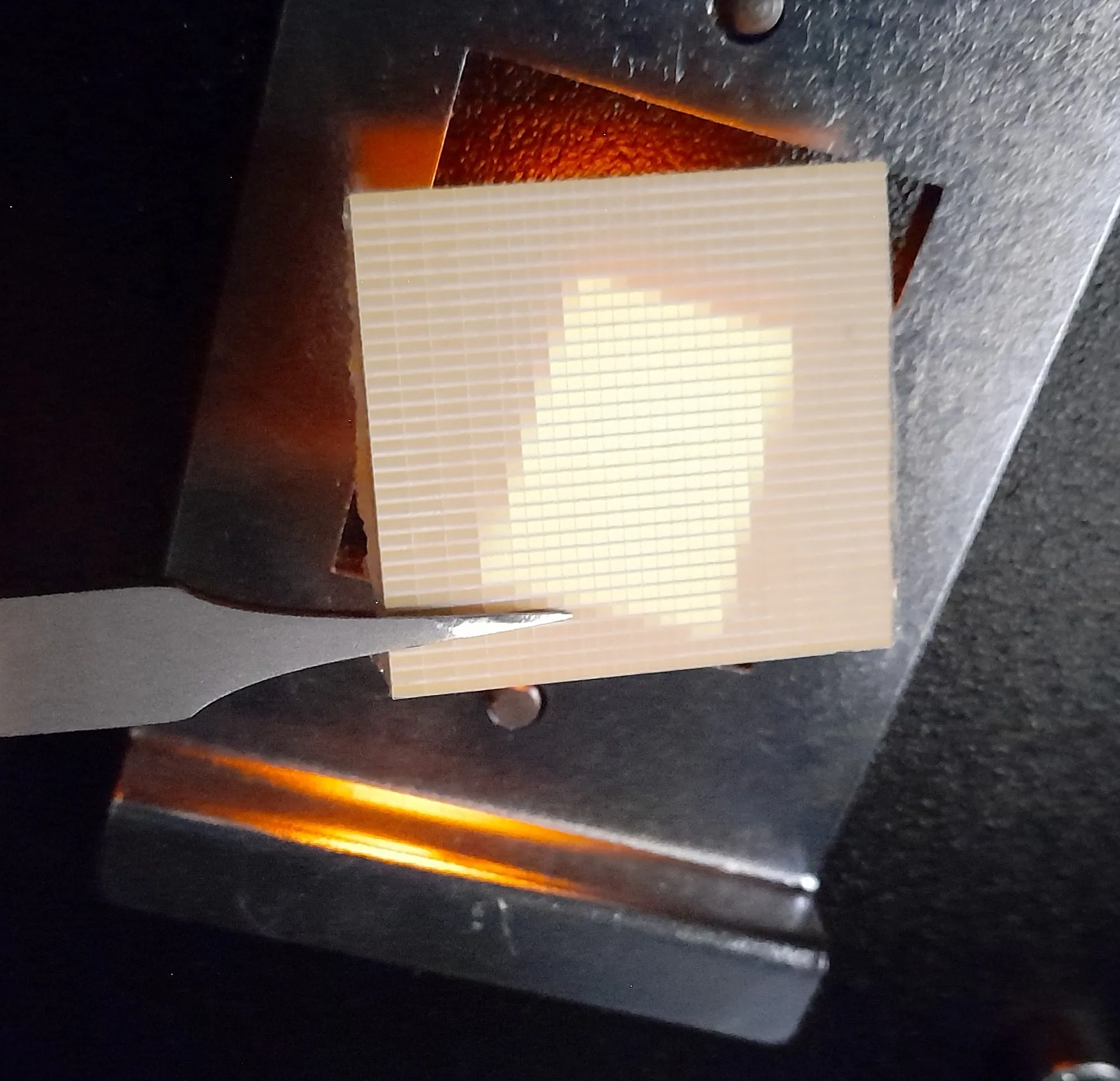
Finally, I tried one more thing: an Electroluminescent (EL) panel. These usually contain copper-doped Zinc sulfide ($ZnS:Cu$) sandwiched between two electrodes. The top electrode is transparent to allow the light to pass through.
If there were a contest for materials not specifically designed for X-ray detection, this would be the clear winner. The light output is strong, and its decay time (how fast the light fades after the beam stops) is excellent. I didn't measure the exact timing, but while the Strontium aluminate glows for seconds, the CFL and LEDs seemed to fade in tenths of a second. The EL foil, however, appeared to turn off almost instantly. While this wasn't a scientific measurement (like nanosecond-scale decay), the EL foil is the winner based on the video.
If there were a contest for materials not specifically designed for X-ray detection, this would be the clear winner. The light output is strong, and its decay time (how fast the light fades after the beam stops) is excellent. I didn't measure the exact timing, but while the Strontium aluminate glows for seconds, the CFL and LEDs seemed to fade in tenths of a second. The EL foil, however, appeared to turn off almost instantly. While this wasn't a scientific measurement (like nanosecond-scale decay), the EL foil is the winner based on the video.
For comparison, I also tested a Gadolinium Oxysulfide (GADOX) ceramic, salvaged from a CT scanner detector module. It's hard to judge from videos shot in the dark, but under slight ambient light, it was clear that GADOX has a higher light yield than the EL foil. Subjectively, I'd estimate the EL foil's performance at about 70% of the GADOX. I don't have data on its speed either, but based on the video, I couldn't distinguish a difference between GADOX and the EL foil.